International Journal
A.I.
A.I.


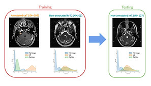
SDC-UDA: Volumetric Unsupervised Domain Adaptation Framework for Slice-Direction Continuous Cross-Modality Medical Image Segmentation, IEEE/CVF Conference on Computer Vision and Pattern Recognition (CVPR), 2023 (Accepted)

A.I.
A.I.


Deep Learning Referral Suggestion and Tumour Discrimination using Explainable Artificial Intelligence applied to Multiparametric MRI, European Radiology, 2023 (Accepted)

A.I.
A.I.


Intelligent Noninvasive Meningioma Grading with a Fully Automatic Segmentation using Interpretable Multiparametric Deep Learning, European Radiology, 2023 (Accepted)

A.I.
A.I.


Digestive Organ Recognition in Video Capsule Endoscopy based on Temporal Segmentation Network, MICCAI, 2022

A.I.
A.I.


CrossMoDA 2021 challenge: Benchmark of cross-modality domain adaptation techniques for vestibular schwannoma and cochlea segmentation, Medical Image Analysis, 2022

A.I.
A.I.


Ultra-thin crystaline silicon-based strain gauges with deep learning algorithms for silent speech interfaces, Nature Communications, 2022

A.I.
A.I.


Small Bowel Detection for Wireless Capsule Endoscopy Using Convolutional Neural Networks with Temporal Filtering, Diagnostics, 2022

A.I.
A.I.


Fully Automatic Quantification of Transient Severe Respiratory Motion Artifact of Gadoxetate Disodium Enhanced MRI during Arterial Phase, Medical Physics, 2022

A.I.
A.I.


M3T: three-dimensional Medical image classifier using Multi-plane and Multi-slice Transformer, Proceedings of the IEEE/CVF Conference on Computer Vision and Pattern Recognition (CVPR), 2022
[Journal Link]

A.I.
A.I.


Importance of CT image normalization in radiomics analysis: prediction of 3-year recurrence-free survival in non-small cell lung cancer. European Radiology, 2022.

A.I.
A.I.


[BlochGAN]
Fat-saturated Image Generation from Multi-contrast MRIs Using Generative Adversarial Networks with Bloch Equation-based Autoencoder Regularization. Medical Image Analysis, Volume 73, October 2021, 102198

A.I.
A.I.


Results of the 2020 fastMRI Challenge for Machine Learning MR Image Reconstruction. -with Facebook AI & NYU. IEEE Transactions on Medical Imaging, (In press)

A.I.
A.I.


Lee, J., Kim, S., Park, I., Eo, T., Hwang, D. (2021). Relevance-CAM: Your Model Already Knows Where to Look. Proceedings of the IEEE/CVF Conference on Computer Vision and Pattern Recognition (CVPR), 2021, pp. 14944-14953

A.I.
A.I.


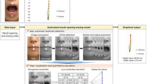
Quantitative analysis of the mouth opening movement of temporomandibular joint disorder patients according to disc position using computer vision: a pilot study. QUANTITATIVE IMAGING IN MEDICINE AND SURGERY , 2021.

A.I.
A.I.


Jun, Y., Shin, H., Eo, T., Hwang, D. (2021). Joint Deep Model-based MR Image and Coil Sensitivity Reconstruction Network (Joint-ICNet) for Fast MRI. Proceedings of the IEEE/CVF Conference on Computer Vision and Pattern Recognition (CVPR), 2021, pp. 5270-5279

A.I.
A.I.


Jun, Y., Shin, H., Eo, T., Kim, T., Hwang, D. (2021). Deep Model-based Magnetic Resonance Parameter Mapping Network (DOPAMINE) for Fast T1 Mapping Using Variable Flip Angle Method. Medical Image Analysis,
70, 102017

A.I.
A.I.


Park, Y.*, Jun, Y.*, Lee, Y. , Han, K., An, C., Ahn, S.**, Hwang, D.**, Lee, S.(2021). Robust Performance of Deep Learning for Automatic Detection and Segmentation of Brain Metastases Using Three-dimensional Black-Blood and Three-dimensional Gradient Echo Imaging. European Radiology, ( In press )

A.I.
A.I.


Shin, H., Lee, J., Eo, T., Jun, Y., Kim, S., Hwang, D. (2020). The Latest Trends in Attention Mechanisms and Their Application in Medical Imaging. Journal of the Korean Society of Radiology, 81(6), 1305-1333.

A.I.
A.I.


Eom, J., Park, I., Kim, S., Jang, H., Hwang, D. (2021). Deep-learned Spike Representations and Sorting via an Ensemble of Auto-encoders. Neural Networks, 134, 131-142.

A.I.
A.I.


Kim, S., Jang, H., Jang, J., Lee, Y., Hwang, D. (2020). Deep-learned Short Tau
Inversion Recovery Imaging Using Multi Contrast Magnetic Resonance Images. Magnetic Resonance in Medicine, 84(6), 2994-3008.

A.I.
A.I.


Eo, T., Shin, H., Jun, Y., Kim, T., Hwang, D. (2020). Accelerating Cartesian MRI by Domain-Transform Manifold Learning in Phase-Encoding Direction. Medical Image Analysis, 63, 101689.

A.I.
A.I.


Jang, H., Bang, K., Jang, J., Hwang, D. (2020). Dynamic Range Expansion Using Cumulative Histogram Learning for High Dynamic Range Image Generation. IEEE Access, 8, 38554-38567.

A.I.
A.I.


Park, I., Eom, J., Jang, H., Kim, S., Park, S., Huh, Y., Hwang, D. (2019). Deep Learning-Based Template Matching Spike Classification for Extracellular Recordings. Applied Sciences, 10(1), 301.

A.I.
A.I.


Hwang, D., Kim, D. (2019). Special Features on Intelligent Imaging and Analysis. Applied Sciences, 9(22), 4804.

A.I.
A.I.


Kim, T., Kim, G., Kim, H., Yoon, H., Kim, T., Jun, Y., Shin, T., Kang, S., Cheon, J., Hwang, D., Min, B., Shim, W. (2019). Megahertz-wave-transmitting conducting polymer electrode for device-to-device integration. Nature Communications, 10(1), 653.

A.I.
A.I.


Jun, Y., Eo, T., Shin, H., Kim, T., Lee, H., Hwang, D. (2019). Parallel Imaging in Time-of-Flight Magnetic Resonance Angiography Using Deep Multi-Stream Convolutional Neural Networks. Magnetic Resonance in Medicine, 81(6), 3840-3853.

A.I.
A.I.


Lee, Y., Kim, S., Suh, J., Hwang, D. (2018). Learning Radiologist’s Step-by-Step Skill for Cervical Spinal Injury Examination: Line drawing, Prevertebral Soft Tissue Thickness Measurement, and Detection of the Swelling in Radiographs. IEEE Access, 6, 55492-55500.

A.I.
A.I.


Jang, H., Bang, K., Jang, J., Hwang, D. (2018). Inverse Tone Mapping Operator Using Sequential Deep Neural Networks Based on Human Visual System. IEEE Access, 6, 52058-52072.

A.I.
A.I.


Kim, S., Bae, W., Masuda, K., Chung, C., and Hwang, D. (2018). Fine-Grain Segmentation of the Intervertebral Discs from MR Spine Images Using Deep Convolutional Neural Networks: BSU-Net. Applied Sciences, 8(9), 1656.

A.I.
A.I.


Jang, J., Jang, H., Eo, T., Bang, K., and Hwang, D. (2018). No-reference Automatic Quality Assessment for Colorfulness-Adjusted, Contrast-Adjusted, and Sharpness-Adjusted Images Using High-Dynamic-Range-Derived Features. Applied Sciences, 8(9), 1688.

A.I.
A.I.


Kim, S., Bae, W., Masuda, K., Chung, C., Hwang, D. (2018). Semi-Automatic Segmentation of Vertebral Bodies in MR images of Human Lumbar Spines. Applied Sciences. 8(9), 1586.

A.I.
A.I.


Oh, D., Kim, S., Park, D., Choi, S., Song, H., Choi, Y., Moon, S., Baek, J., Hwang, D. (2018). Correction of severe beam-hardening artifacts via a high-order linearization function using a prior-image-based parameter selection method. Medical Physics, 45(9), 4133-4144.

A.I.
A.I.


Jun, Y., Eo, T., Kim, T., Shin, H., Hwang, D.*, Bae, S., Park, Y., Lee, H., Choi, B., Ahn, S. (2018). Deep-learned 3D black-blood imaging using automatic labelling technique and 3D convolutional neural networks for detecting metastatic brain tumors. Scientific Reports, 8: 9450.

A.I.
A.I.


Eo, T., Jun, Y., Kim, T., Jang, J., Lee, H., & Hwang, D. (2018). KIKI-net: Cross-Domain Convolutional Neural Networks for Reconstructing Undersampled Magnetic Resonance Images. Magnetic Resonance in Medicine, 80(5), 2188-2201.

A.I.
A.I.


Jang, J., Bang, K., Jang, H., & Hwang, D. (2018). Quality Evaluation of No-reference MR Images Using Multidirectional Filters and Image Statistics. Magnetic Resonance in Medicine, 80(3), 914-924.

A.I.
A.I.


Lee, Y., & Hwang, D. (2018). Periodicity-based nonlocal-means denoising method for electrocardiography in low SNR non-white noisy conditions. Biomedical Signal Processing and Control, 39, 284-293.

A.I.
A.I.


Kim, Y., Oh, D., & Hwang, D. (2017). Small-scale noise-like moiré pattern caused by detector sensitivity inhomogeneity in computed tomography. Optics Express, 25(22), 27127-27145.

A.I.
A.I.


Song, K. I., Chu, J. U., Park, S. E., Hwang, D., & Youn, I. (2017). Ankle-Angle Estimation from Blind Source Separated Afferent Activity in the Sciatic Nerve for Closed-Loop Functional Neuromuscular Stimulation System. IEEE Transactions on Biomedical Engineering, 64(4), 834-843.

A.I.
A.I.
-contrast%20magnetic.png)

Eo, T., Kim, T., Jun, Y., Lee, H., Ahn, S. S., Kim, D. H., & Hwang, D. (2017). High‐SNR multiple T2 (*)‐contrast magnetic resonance imaging using a robust denoising method based on tissue characteristics. Journal of Magnetic Resonance Imaging, 45(6), 1835-1845.

A.I.
A.I.


Hwang, D., Kim, S., Abeydeera, N. A., Statum, S., Masuda, K., Chung, C. B., ... & Bae, W. C. (2016). Quantitative magnetic resonance imaging of the lumbar intervertebral discs. Quantitative Imaging in Medicine and Surgery, 6(6), 744-755.

A.I.
A.I.


Park, H. S., Hwang, D., & Seo, J. K. (2016). Metal artifact reduction for polychromatic x-ray CT based on a beam-hardening corrector. IEEE transactions on medical imaging, 35(2), 480-487.

A.I.
A.I.


Kim, S., & Hwang, D. (2015). Murmur-adaptive compression technique for phonocardiogram signals. Electronics Letters, 52(3), 183-184.

A.I.
A.I.


Nam, Y., Lee, J., Hwang, D., & Kim, D. H. (2015). Improved estimation of myelin water fraction using complex model fitting. NeuroImage, 116, 214-221.

A.I.
A.I.


Park, S. E., Song, K. I., Suh, J. K. F., Hwang, D., & Youn, I. (2015). A time-course study of behavioral and electrophysiological characteristics in a mouse model of different stages of Parkinson's disease using 6-hydroxydopamine. Behavioural brain research, 284, 153-157.

A.I.
A.I.


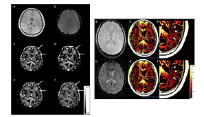
Gho, S. M., Liu, C., Li, W., Jang, U., Kim, E. Y., Hwang, D., & Kim, D. H. (2014). Susceptibility map‐weighted imaging (SMWI) for neuroimaging. Magnetic resonance in medicine, 72(2), 337-346.

A.I.
A.I.


Kim, Y., Baek, J., & Hwang, D. (2014). Ring artifact correction using detector line-ratios in computed tomography. Optics express, 22(11), 13380-13392.

A.I.
A.I.


Hwang, D., & Zeng, G. L. (2014). Special issue on medical imaging. Biomedical Engineering Letters, 4(1), 1-2.

A.I.
A.I.


Chu, J. U., Song, K. I., Shon, A., Han, S., Lee, S. H., Kang, J. Y., ... & Youn, I. (2013). Feedback control of electrode offset voltage during functional electrical stimulation. Journal of neuroscience methods, 218(1), 55-71.

A.I.
A.I.


Kwon, O. I., Woo, E. J., Du, Y. P., & Hwang, D. (2013). A tissue-relaxation-dependent neighboring method for robust mapping of the myelin water fraction. NeuroImage, 74, 12-21.

A.I.
A.I.


Chu, J. U., Song, K. I., Han, S., Lee, S. H., Kang, J. Y., Hwang, D., ... & Youn, I. (2013). Gait phase detection from sciatic nerve recordings in functional electrical stimulation systems for foot drop correction. Physiological measurement, 34(5), 541.

A.I.
A.I.


Jang, U., Nam, Y., Kim, D. H., & Hwang, D. (2013). Improvement of the SNR and resolution of susceptibility-weighted venography by model-based multi-echo denoising. Neuroimage, 70, 308-316.

A.I.
A.I.


Kang, B., Choi, O., Kim, J. D., & Hwang, D. (2013). Noise reduction in magnetic resonance images using adaptive non-local means filtering. Electronics Letters, 49(5), 324-326.

A.I.
A.I.


Chu, J. U., Song, K. I., Han, S., Lee, S. H., Kim, J., Kang, J. Y., ... & Youn, I. (2012). Improvement of signal-to-interference ratio and signal-to-noise ratio in nerve cuff electrode systems. Physiological measurement, 33(6), 943.

A.I.
A.I.


Lee, Y. J., Lee, P. J., Kim, K. S., Park, W., Kim, K. D., Hwang, D., & Lee, J. W. (2012). Toothbrushing region detection using three-axis accelerometer and magnetic sensor. IEEE Transactions on Biomedical Engineering, 59(3), 872-881.

A.I.
A.I.
_contrast_MR_.png)

Jang, U., & Hwang, D. (2012). High‐quality multiple T2 (*) contrast MR images from low‐quality multi‐echo images using temporal‐domain denoising methods. Medical physics, 39(1), 468-474.

A.I.
A.I.


Hwang, D., Lee, J. W., & Zeng, G. L. (2011). SPECT reconstruction with sub‐sinogram acquisitions. International Journal of Imaging Systems and Technology, 21(3), 247-252.

A.I.
A.I.


Hwang, D., Chung, H., Nam, Y., Du, Y. P., & Jang, U. (2011). Robust mapping of the myelin water fraction in the presence of noise: synergic combination of anisotropic diffusion filter and spatially regularized nonnegative least squares algorithm. Journal of Magnetic Resonance Imaging, 34(1), 189-195.

A.I.
A.I.


Hwang, D., Kim, D. H., & Du, Y. P. (2010). In vivo multi-slice mapping of myelin water content using T2* decay. NeuroImage, 52(1), 198-204.

A.I.
A.I.


Hwang, D., & Du, Y. P. (2009). Improved myelin water quantification using spatially regularized non‐negative least squares algorithm. Journal of Magnetic Resonance Imaging, 30(1), 203-208.

A.I.
A.I.


Du, Y. P., Chu, R., Hwang, D., Brown, M. S., Kleinschmidt‐DeMasters, B. K., Singel, D., & Simon, J. H. (2007). Fast multislice mapping of the myelin water fraction using multicompartment analysis of T decay at 3T: A preliminary postmortem study. Magnetic Resonance in Medicine, 58(5), 865-870.

A.I.
A.I.


Nagarajan, S. S., Portniaguine, O., Hwang, D., Johnson, C., & Sekihara, K. (2006). Controlled support MEG imaging. NeuroImage, 33(3), 878-885.

A.I.
A.I.


Hwang, D., & Zeng, G. L. (2005). Convergence study of an accelerated ML-EM algorithm using bigger step size. Physics in medicine and biology, 51(2), 237.

A.I.
A.I.


Hwang, D., & Zeng, G. L. (2005). Reduction of noise amplification in SPECT using smaller detector bin size. IEEE transactions on nuclear science, 52(5), 1417-1427.

A.I.
A.I.


Hwang, D., & Zeng, G. L. (2005). A new simple iterative reconstruction algorithm for SPECT transmission measurement. Medical physics, 32(7), 2312-2319.

U.S. Patents
11. US 10,168,405 B2 (2019/01/01), METHOD AND APPARATUS FOR QUANTIFYING PROPERTIES OF AN OBJECT THROUGH MAGNETIC
RESONANCE IMAGING (MRI)
10. US 9,989,609 B2 (2018/06/05), METHOD AND APPARATUS FOR ADJUSTING THE PARAMETERS OF A MAGNETIC RESONANCE IMAGE
9. US 9,977,109 B2 (2018/05/22), MAGNETIC RESONANCE IMAGING APPARATUS AND OPERATING METHOD FOR THE SAME
8. US 9,928,576 B2 (2018/03/27), DENOISING METHOD AND APPARATUS FOR MULTI-CONTRAST MRI.
7. US 9,759,796 B2 (2017/09/12), METHOD AND APPARATUS FOR ELIMINATING NOISE IN MAGNETIC RESONANCE IMAGES.
6. [Germany] Patent Granted: METHOD AND APPARATUS FOR ELIMINATING NOISE IN MAGNETIC RESONANCE IMAGES.
5. [France] Patent Granted: METHOD AND APPARATUS FOR ELIMINATING NOISE IN MAGNETIC RESONANCE IMAGES.
4. [UK] Patent Granted: METHOD AND APPARATUS FOR ELIMINATING NOISE IN MAGNETIC RESONANCE IMAGES.
3. US 9,506,896 B2 (2016/11/29), METHOD AND APPARATUS FOR DETECTING AN ENVELOPE FOR ULTRASONIC SIGNALS.
2. US 9,241,671 B2 (2016/01/26), APPARATUS AND METHOD FOR REMOVING NOISE FROM BIOSIGNALS.
1. US 9,119,546 B2 (2015/09/01), R-PEAK DETECTION APPARATUS AND CONTROL METHOD THEREOF.
Domestic Patents
49. 컴퓨터 단층촬영 영상 재구성 장치 및 방법
48. 자기 공명 영상 장치 및 자기 공명 영상 장치의 재촬영 제어 방법
47. 심전도 신호를 이용한 사용자 인증 방법 및 장치
46. 자기 공명 영상 장치 및 자기 공명 영상 장치의 재촬영 제어 방법
45. 뇌종양 판별 영상 생성 학습 장치 및 방법과 뇌종양 판별 영상 생성 장치 및 방법 그리고 이에 관한 기록 매체
44. 의료 영상에 기반하여 상태를 진단하는 장치 및 방법
43. 언더샘플링된 자기 공명 영상의 재구성 장치 및 방법
42. 화질 평가 장치 및 방법과 이에 관한 기록매체
41. 합성곱 신경망 딥 러닝 기반 경추 연조직의 측정 방법
40. 후처리 기반 자기공명영상의 파라미터 조정 방법 및 장치
39. 컴퓨터 단층 촬영 영상의 아티팩트를 보정하는 방법 및 장치
38. 단일 엑스레이를 이용한 병변 위치측정 기술
37. 움직임 추적을 위한 관심영역 설정 장치 및 방법과 그 기록매체
36. 자기 공명 영상 장치에서 위상 펼침 방법 및 그 방법을 사용하는 자기 공명 영상 장치
35. 자기 공명 영상을 통해 대상체의 속성을 정량화하기 위한 방법 및 장치
34. 복소수 신호 모델링을 이용한 다중 자기공명영상에서의 위상 복원 방법
33. 움직임 추적을 위한 ROI 설정 방법
32. 자기공명영상의 파라미터 조정 방법 및 장치
31. 자기 공명 영상 장치 및 그 동작방법
30. 초음파 신호의 포락선 검출 방법 및 그 장치
29. 다중 대조도 자기공명영상에서 잡음 제거 방법 및 장치
28. 초음파 신호의 포락선 검출 방법 및 그 장치
27. 종양 추적 방법 및 그 장치
26. 자기 공명 영상 장치 및 그 동작방법
25. 후처리 기반 자기공명영상의 파라미터 조정 방법 및 장치
24. 자기공명영상의 파라미터 조정 방법 및 장치
23. 자기공명영상 잡음 제거 방법 및 장치
22. 종양특성에 기반한 종양 추적 방법 및 그 장치
21. 자기공명영상 잡음 제거 방법 및 장치
20. 다중 대조도 자기공명영상에서 잡음 제거 방법 및 장치
19. 초음파 신호의 포락선 검출 방법 및 그 장치
18. 자기 공명 영상 장치 및 그 동작방법
17. R 피크 검출을 위한 심전도 장치 및 방법
16. 듀얼 촬영 방식 컴퓨터 단층 촬영장치 및 이를 이용한 컴퓨터 단층 촬영방법
15. 듀얼 프로브가 구비된 초음파 촬영장치 및 이를 이용한 초음파 촬영방법
14. ECG 신호를 이용한 환자의 청진위치정보 획득 장치 및 이를 이용한 환자의 청진위치정보 획득 방법
13. 생체 신호의 노이즈 제거 장치 및 방법
12. 자기공명영상 잡음 제거 방법 및 장치
11. R - 피크 검출 장치 및 그 제어 방법
10. 생체신호를 이용한 초음파 진단 시스템 및 진단 방법
09. ECG 신호 검출 시스템 및 방법
08. 보정용 팬텀을 이용한 CT 영상 획득장치 및 획득방법
07. 심전도 신호를 이용한 사용자 인증 방법 및 장치
06. 생체 신호의 노이즈 제거 장치 및 방법
05. R - 피크 검출 장치 및 그 제어 방법
04. 의료용 융합영상 획득장치 및 획득방법
03. 실시간 단층 영상 생성장치, 생성방법 및 실시간 단층 영상을 이용한 의료장치
02. 단층 영상 생성 장치 및 방법
01. 의료용 융합영상 획득장치 및 획득방법
