

Joint Research Laboratory Yonsei-UCSD


MAI-LAB @


History of JRL Yonsei-UCSD
-
2015, Started research supervisor communication
-
2016, graduate student participation
-
2017, Yonsei-UCSD Joint Research Laboratory Agreement
-
2017, Published results of research to international journals
-
2018, Started deep research interchange and published SCI papers
-
2019, Visited UCSD
-
2019, Installed AI (Artificial Intelligence) program developed from Yonsei Univ. in UCSD laboratory
-
2019, Submitted joint research results to RSNA (Radiological Society of North America)
-
2019, Research meeting in Montreal, Canada
Disc and Vertebral Body Segmentation in Lumbar Spine MR


-
Intervertebral disc segmentation in MR images is challenging owing to their complex shapes and non-uniform intensity.
-
This study introduces a robust deep-learning segmentation network which can successfully segment intervertebral discs with complex boundaries.

Joint Research Laboratory Yonsei-Martinos Center
Athinoula A. Martinos Center for Biomedical Imaging
Massachusetts General Hospital / Harvard Medical School
MAI-LAB @


History of JRL Yonsei-Martinos Center
-
2022, Yohan Jun, an MAI-LAB alumnus, joined the Martinos Center as a Postdoctoral Researcher
-
2024, Yohan Jun was promoted to instructor in Radiology at Harvard Medical School
-
2025, Jinho Joo, a Ph.D. student at MAI-LAB, began collaborative research with the Martinos Center
-
2026, MAI-LAB and the Martinos Center established the Yonsei-Martinos Center Joint Research Laboratory (JRL)
-
2026, Yohan Jun was appointed Assistant Professor at Korea University
-
2026, Jinho Joo joined the Martions Center as a Visiting Researcher through the JRL collaboration
-
Vertebral body segmentation on MR images provides clinically useful information including quantitative biomarkers, volume, and shape.
-
This study proposes a semi-automatic algorithm for the segmentation of vertebral bodies in magnetic resonance images of the human lumbar spine.
-
Our method achieved a 90% dice similarity coefficient and significantly reduces the user’s role.
Knee Cartilage Segmentation in Knee MR
-
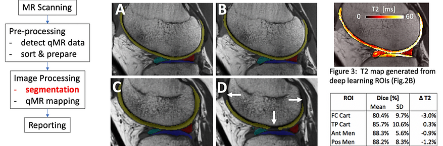
In a translational diagnostic workflow of the knee MRI involving quantitative sequences, there is a need for segmentation in order to evaluate specific regions.
-
In the knee, articular cartilage and menisci are among those frequently segmented, to determine quantitative MR (qMR) values such as T2.
-
This study demonstrates an application for deep learning (DL)-based automated segmentation of cartilage and menisci.


